What is an ultrasound?
Ultrasonography is an imaging method which uses sound waves to produce images of organs and tissues within the body. It is based on the same principles involved in sonar used by ships and fisherman.
Sound waves are directed at organs and tissues of the body. When these waves encounter tissues, they bounce back and produce echoes. These echoes are manipulated by the ultrasound machine to produce images of the body.

How we perform our ultrasounds
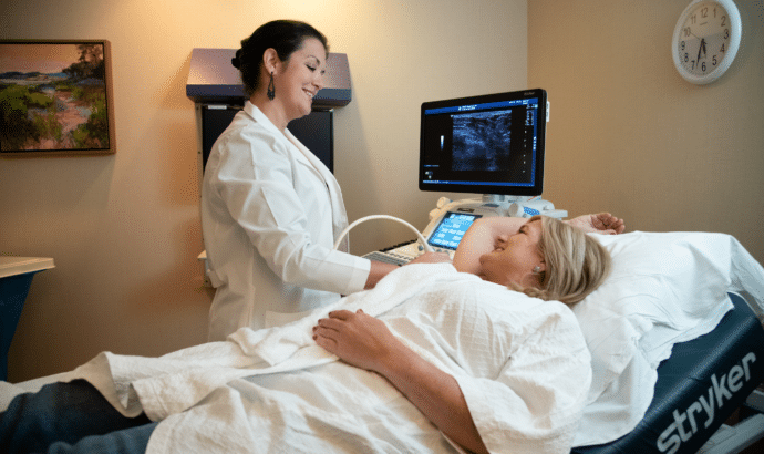
An ultrasound exam is performed by a sonographer, a technician trained in ultrasound imaging. During the procedure, the technician presses a small hand-held device, called a transducer, against your skin. The transducer generates and receives sound waves. The technologist moves the transducer over the skin overlying the tissues or organs in question. Sound waves generated by the transducer bounce off tissues and organs within your body and reflected sound waves are recorded by the transducer. Differing organs and tissues produce unique echoes or sound reflections. This information is then sent to a computer which produces detailed images of the organ or body part in question-based on the unique patterns created by the reflected sound.
Ultrasound is a valuable tool utilized by your doctor. It is used by physicians in the assessment of symptoms such as pain, swelling and infection. Ultrasonography is utilized in examining many of the body’s organs including the: heart, liver, gallbladder, spleen, kidneys, pancreas, uterus, ovaries, bladder, thyroid, parathyroid, scrotum, breasts, and blood vessels. Additionally, it is vital in the assessment of pregnancy and the unborn fetus. Other uses include guidance for biopsies, aspirations and drainages.
Types of ultrasounds
- Echocardiogram
- Thyroid
- Abdomen/Pelvis
- Kidneys
- Female Organs-Transvaginal
- Blood Vessels-Carotids, Abdominal Aorta
- Infant Hip
- Head/Neck-Soft Tissues
- Chest/Mediastinum
- Gallbladder
- Bladder
- Scrotum
- Venous Doppler-Extremities
- Ultrasound Procedures
- Guided Biopsy of the Thyroid Gland
- Thoracentesis
- Guided Biopsy of Solid Organs
- Paracentesis